Swift & Accurate Ultrasound
Accurate Soft tissue imaging Through Sound Waves
Ultrasound uses a transducer (probe) to create sound waves and produce images of the body’s internal structures. It does not use ionizing radiation, has no known harmful effects, and provides detailed views of soft tissues that may not be visible on X-rays.
Ultrasound is commonly used to investigate unexplained pain, swelling, or infection. It can also guide needle biopsies or assess conditions related to blood flow.

Ultrasound Common Applications
Soft Tissue & Organs
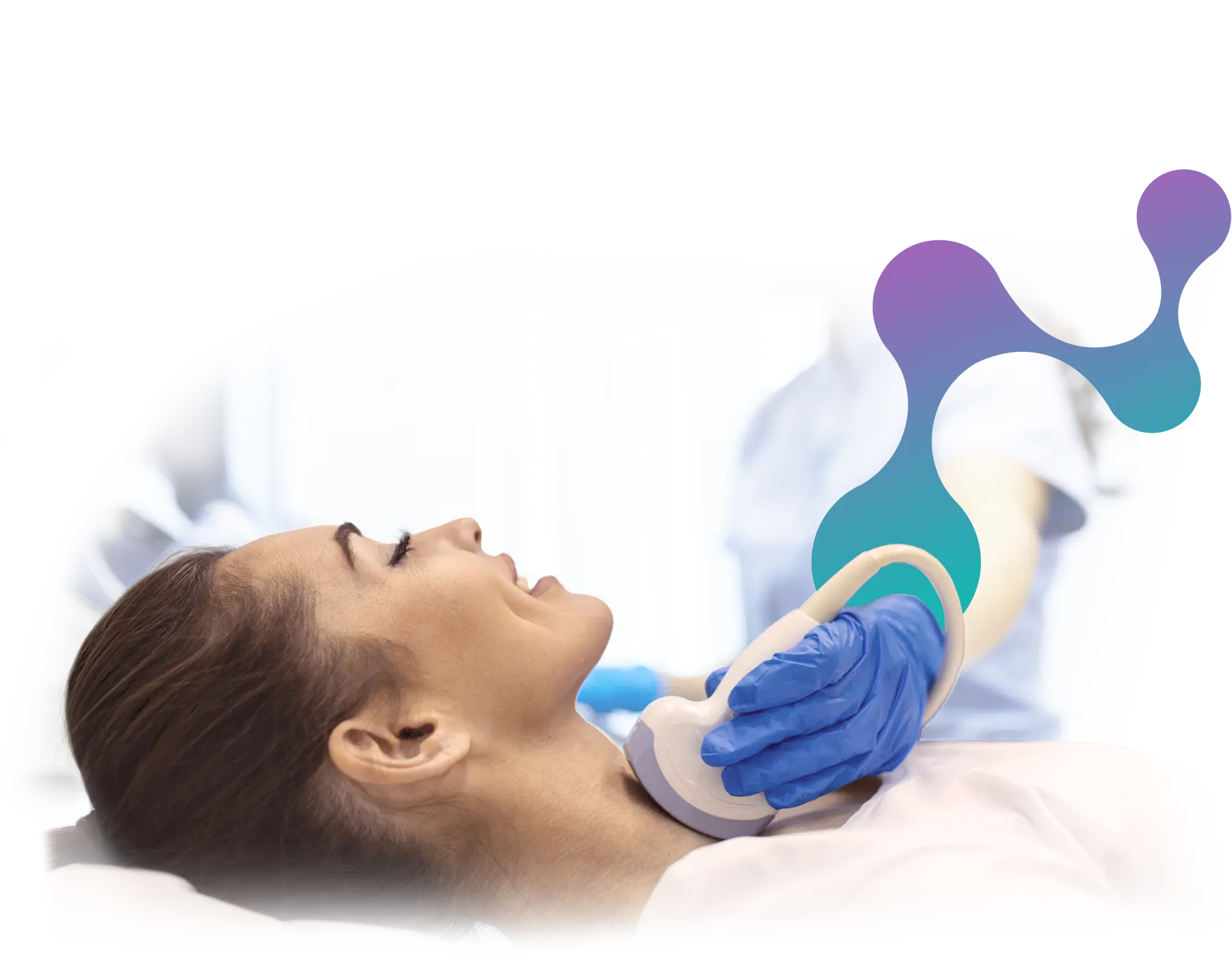
Thyroid
Meticulous screening and detailed analysis of thyroid nodules, screening for Thyroid cancer and Thyroiditis. ATA (American Thyroid association) classification of thyroid nodules, and cancer risk analysis. Patient friendly procedures including ultrasound guided Fine Needle Aspiration Biopsies (FNAB), core biopsies, ultrasound guided cyst aspirations, ultrasound guided alcohol ablations of cysts and neck masses.
Neck
Systematic analysis of cervical lymph nodes, salivary glands and neck masses, with ultrasound staging of neck neoplasms, and ultrasound guided biopsy of suspicious lesions.
Abdominal
Abdominal ultrasound is a non-invasive medical imaging technique that provides valuable insights into internal organs, aiding in diagnosing conditions like gallstones, tumors, and inflammation quickly and safely.
Renal Tracts
Renal tract ultrasound is a painless, radiation-free imaging method that assesses kidney and urinary system health, helping diagnose issues like kidney stones, infections, and structural abnormalities accurately and efficiently.
Pelvic
Pelvic ultrasound is a non-invasive diagnostic tool, particularly beneficial for examining reproductive and urinary organs in both men and women, aiding in detecting issues such as cysts, tumors, and pregnancy detection.
Musculoskeletal & Peripheral
MSK / Joints
Musculoskeletal (MSK) ultrasound is a precise imaging technique that enables the visualization of muscles, tendons, and joints, facilitating accurate diagnosis and treatment of musculoskeletal conditions like injuries and arthritis.
Scrotal
Non invasive assessment of the Testes, epididymi, scrotal content and Iguinal regions.
Vascular
Vascular Sections
Carotid Doppler
Peripheral
- Arterial Doppler
- Venous Doppler
Interventional
Ultrasound Guided
- Biopsies
- Aspirations
- Drainages
- Diagnostic and therapeutic joint injections.
- More about Interventional Radiology >
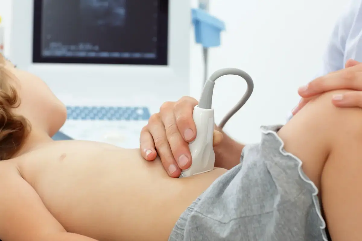
Paediatric
Paediatric Imaging
Pediatric ultrasound is a safe and non-invasive imaging technique tailored for children, helping diagnose various conditions in young patients, from congenital anomalies to infections, without exposing them to radiation.
Neonatal
- Hip
- Cranial
- Abdominal e.g. Gastro-oesophageal reflux screening
Ultrasound Preparation
Abdominal
Upper Abdomen (liver, gallbladder, pancreas, spleen), kidneys, or aorta. Eat a light, low-fat dinner the night before and fast for 8 hours before your exam. You may drink water, juice, or black tea/coffee.
Pelvis
You will need a full bladder. Drink 4–6 glasses of water or other liquids, finishing 1 hour before your exam. Do not empty your bladder beforehand.
Thyroid, limbs, or other areas
No preparation required.
During the Examination
Throughout the non-invasive procedure Dr Berk will explain the procedure and ensure the patient is as comfortable as possible.
You will lie on the exam bed while the examiner uses a probe to perform the scan. A warm gel will be applied to your skin to improve contact. You may be asked to hold your breath or roll onto your side to help obtain clear images.